A better look
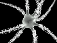
Despite the fact that scientists are able to look inside the brain using a variety of live imaging techniques, their ability to visualize individual neurons in living animals is very limited. Thus our understanding of what is really going on during learning and disease, for example, is also incomplete. A new technique developed by Dr. Mark Schnitzer at Stanford University, however, may change that, by letting scientists look at individual neurons in areas of the brain not accessible by traditional imaging tools.
Known as time-lapse microsendoscopy, this new technique allowed Dr. Schnitzer’s group to observe the same population of neurons in the hippocampus, a brain area involved in learning and memory, over the course of seven weeks in live animals. While the study focused on the effects of gliomas, a type of tumor that originates in the brain, the technique could also be used to monitor changes associated with diseases such as cancer, stroke, or Parkinson’s disease.While microendoscopy is currently being used to diagnose certain forms of cancer, this is the first reported use of microendoscopy over a long period of time. This unique development may help scientists to understand how diseases in the brain and other organs progress, which may ultimately lead to better therapies.
Find out more about how time-lapse microendoscopy in the brain works from this interview with Dr. Schnitzer.
You can also read this blog on my blog site.

