This student story was published as part of the 2026 NASW Perlman Virtual Mentoring Program organized by the NASW Education Committee, providing science journalism experience for undergraduate and graduate students.
Story by Leighana Luther
Mentored and edited by Erin Digitale
In classic educational cartoons like Osmosis Jones and Magic School Bus, miniaturized fictional characters visit the bustling realm inside the human body. These stories are unique because they show viewers what they can’t see in their own bodies: a multitude of organs, tissues, and fluids performing specific actions that work to keep the body alive and functioning.
If the gastrointestinal (GI) tract is a bus route, each bus stop along the route from mouth to anus has a specialized function and environment. Our knowledge of the environment of each of these stops is limited because gastrointestinal research often relies on stool samples.
“You're not getting a picture of the whole microbiome, you're only getting a snapshot of part of it,” said Roujheen Sabetan, a research coordinator at Stanford University School of Medicine. In other words, stool samples represent the very last bus stop, not the entire route.
Tiny explorers, in the form of swallowable devices, have made these previously mysterious areas accessible. This kind of technology is a hot area of research. There are devices being studied that aim to collect intestinal fluid, image the gut, and gather metrics such as pH, temperature, gas, and pressure. While it might sound strange to swallow a medical device, these devices can provide useful data about distant areas of the gut.
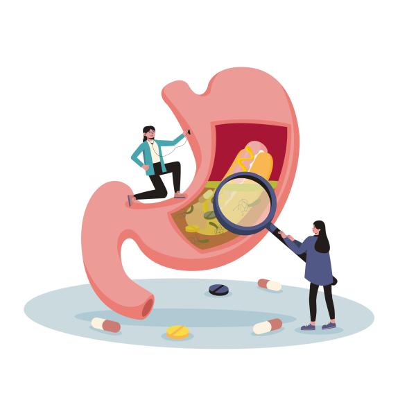
A person holding a magnifying glass over a stomach. Credit: Unsplash.
What’s going on inside the gut?
In the mouth, saliva swirls, initiating digestion. Among the blush-colored canyons and valleys of the coiled stomach folds, acid helps to break down food. In the small intestine, these coiled folds are close together, representing the vast surface area the intestines contain. If the GI tract could be stretched out and measured, it would be about 30 feet long. In the last section, the muscles of the large intestine contract and release to move waste along.
Procedures like endoscopy and colonoscopy reach a few feet into the GI tract from the mouth or anus, respectively, but reaching the depths of the GI tract near the middle is much more difficult. Invasive options like surgery can give us a clear view of the physical structure of these hard-to-reach spots, but can’t show us how they operate in an ordinary day.
How do these devices work?
Dr. Asma Khapra, a gastroenterologist specializing in women’s digestive health and inflammatory bowel disease, frequently uses a device called a camera pill to view the small intestines of her patients. “For an upper endoscopy, you get to the top of the small bowel and you can't go further, and with a colonoscopy you get to the bottom of the small intestine,” said Khapra. “The small bowel is very difficult to visualize with a scope, so that’s why the video capsule technology is used.”
The notion of downing an inedible item might be unnerving, but these devices are specifically designed to be safe to swallow, in contrast to objects that people (mostly children) accidentally swallow such as coins, beads, or small electronics. However, it can still be tricky for some people.
“It’s like a big horse pill. You know, occasionally people will be like, ‘I can’t swallow it, like I just, I can’t do that.’ But, you know, the majority of people can swallow it,” said Khapra.
The resulting video helps her look for active bleeding that may be causing iron-deficiency anemia or ulcers that may be suggestive of Crohn’s disease.
The camera pill entered the clinical landscape back in 2001, but new devices still in experimental phases continue to pop up in research.
One such new device is the CapScan, an experimental capsule that collects fluid from the GI tract. About the size and shape of a Tylenol, the CapScan cleverly slips a 30 cm collection tube into the gut. The flexible tube is packed into the small capsule, and as it moves through the gut, the outer coating dissolves. The tube slowly begins to unravel, and as it’s exposed, it pulls the surrounding liquid inside, much like how a paper towel soaks up a spill. This gathers samples from multiple locations within the gut.
Patients collect the device using a collection kit provided by the researchers that contains materials such as gloves, biohazard bags, and stool collection containers. Sabetan, who coordinates CapScan research at Stanford, said, “For most of [the patients], that's their least favorite part, just because most people don't want to be sorting through their stool.”
Once the device is returned to researchers, they use the fluid to analyze the patient’s gut microbiome, or the unique collection of microorganisms every individual has inside of them. Sabetan said, “We're starting to look for patterns and things that would relate the symptoms to what the microbiome looks like.” They collect samples from healthy patients, as well as those with all kinds of GI distress, including conditions like functional bloating and irritable bowel syndrome.
What’s next?
More research needs to be conducted to confidently link an individual’s microbiome with specific conditions. Although they are still experimental, devices like this may one day be available to offer personalized assessments and guide diagnosis or treatment of GI conditions. “There's still so much to learn that it's a very fun field to be doing research on,” said Sabetan.
Main Header Image Caption: Woman holding half-full glass and white medicine pill. Credit: Pexels.

Leighana Luther is a freelance science writer and graduate student in the Science Writing MA program at Johns Hopkins University.
Erin Digitale is a senior science writer at Stanford Medicine.
The NASW Perlman Virtual Mentoring program is named for longtime science writer and past NASW President David Perlman. Dave, who died in 2020 at the age of 101 only three years after his retirement from the San Francisco Chronicle, was a mentor to countless members of the science writing community and always made time for kind and supportive words, especially for early career writers.
You can contact the NASW Education Committee at education@nasw.org. Thank you to the many NASW member volunteers who lead our #SciWriStudent programming year after year.
Founded in 1934 with a mission to fight for the free flow of science news, NASW is an organization of ~ 2,300 professional journalists, authors, editors, producers, public information officers, students and people who write and produce material intended to inform the public about science, health, engineering, and technology. To learn more, visit www.nasw.org